Tuesday, January 30, 2018

Shock Value

Shock Value: Electrifying Hunting Techniques of Electric Eels

Electrophorus electricus


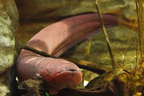
Electric eels don’t look like fierce hunters.  They have tiny eyes, skinny bodies, and dopey-looking expressions.  However, don’t be fooled.  These fish(they aren’t really eels), pack enough electric force to incapacitate their prey and send much larger animals jerking hastily away. 

The electric eel uses three specialised organs to produce electricity: Sach’s organ, the main electric organ, and Hunt’s organ.  These take up the vast majority of the eel’s body 1.  The main electric organ produces the most of the fish’s strong electric charge, giving it the ability to stun prey and defend itself from predators.  Sach’s Organ generates continual low electric currents round the fish, which allows it to sense surrounding objects based on how they interact with the field 2. Hunt’s organ is somewhat more mysterious, and is believed to help both other organs hunting and navigation 2.  These structures, along with pressure-sensing hairs along the fish's body, allow E. electricus to hunt in the muddy river bottoms they calls home.  
Fig.1: Structure and location of electric organs E. electricus.  Because of their similarity in structure and protein composition to muscle, the electricity-producing organs of these fish are believed to have evolved from muscle tissue.

Fig.2 : Electrolocation is the process of using electric fields to sense the surrounding environment.  The field generated by Sach’s organ is interrupted by objects in its way.  Electric eels can sense the charges in field and use them to determine if they are passing by a large immobile rock or a small mobile fish.


To generate electricity, the eel has developed stacks of specialized cells called electrocytes3.  One one side of the electrocyte is ruffled and rich in a protein pump called the sodium-potassium ATPase.  This constantly pumps three positively charged sodium ions out of the cell and pumps two positively charged potassium in, giving the interior of the cell a negative charge versus the outside of the cell.  When the nerves connecting the opposite side of the cell fire, they release a compound called acetylcholine.  Acetylcholine binds another ion channel on the electrocytes, which opens and allows large amounts of sodium to rush in and small amounts of potassium to rush out.  In this stimulated state, one side of the cell is negative and the other is positive.  The charge difference within the cell generates an eel’s electric power.  
 
Figure 3: Electrocytes at rest(L) and stimulated(R).  In the resting state, Na+/K+-ATPase maintains a net negative charge inside the cell.  When neurons secrete acetylcholine into the space between nerves and electrocytes, channels in the plasma membrane open on one side of the cell and allow large amounts of sodium to rush in.  This gives that side of the cell a positive charge.

Each electrocyte can produce around 0.15V, but stacked and lined up in rows they can produce up to 600V 4.  Discharging the electrocytes all at once creates a strong electrical current from the eel’s head to tail.  If anything gets in the way of that field it receives a sharp shock. 
Figure 4:When a large number of electrocytes are lined up and charged, their combined positive and negative charges generate a stronger voltage.


E. electricus uses it's electric organs in a unique hunting method.  When an electric eel senses movement nearby it will first generate two brief electrical shocks4.  The shocks cause nearby fish or other creatures’ muscles to twitch.  Then, alerted to its target's location, the eel will grab its prey.  With the surprised creature held in its jaws, it curls into a circle and pins the prey between its head and tail.  Its prey is trapped there in the strongest part of the field, as the eel generates shock after shock in quick succession.  The prey’s muscles are quickly overstimulated and fatigued by the electric shocks.  While the prey is in this stunned state E. electricus wolfs down its meal4. 
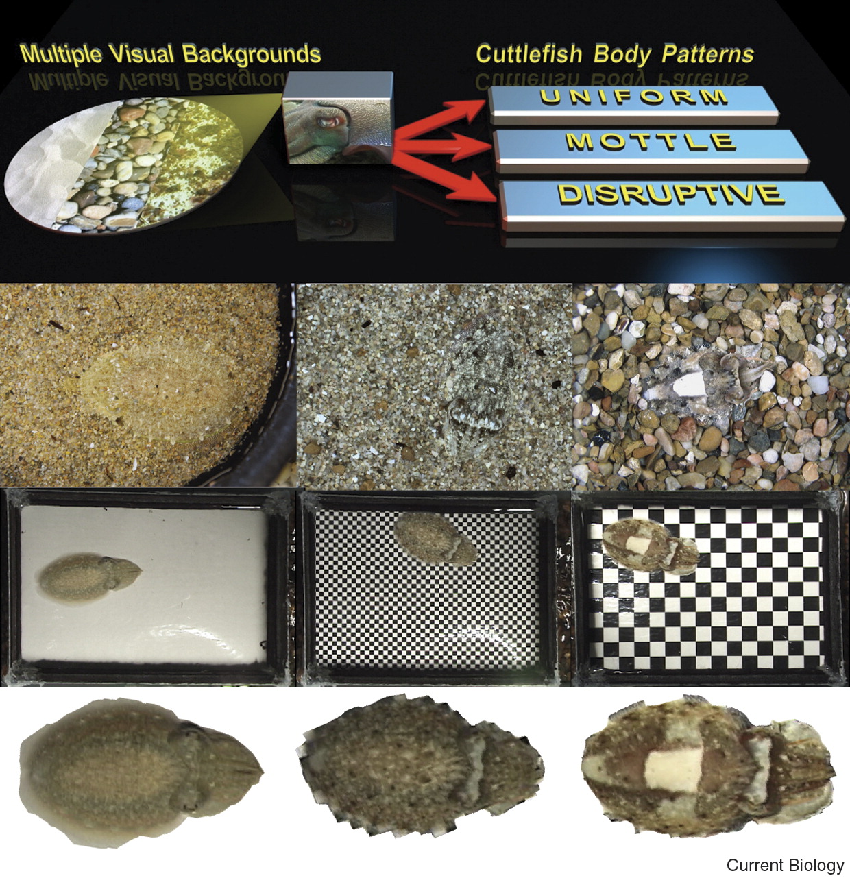
Fig. 5: Electric eel hunting behaviors.  Electrophorus electricus curl into a circle to put their prey in the strongest section of the electric field before emitting a series of rapid shocks.  The high voltage causes they prey's muscles to contract, until they are temporarily unable to move.


Video 1: In the video above, the shocks an electric eel generates while hunting is shown by flashing red light. 

Adult electric eels can not only produce a powerful dose of electricity, they can control the amount of electricity they discharge5.  This allows them to save energy if they’re hunting a small crab versus defending themselves from a large fish or bird.  Although they primarily have been observed shocking other creatures in the water, electric eels have also been observed jumping out of the water to shock alligators, horses, and humans.  One scientist confirmed this behavior by allowing electric eels to repeatedly shock his arm, while he measured the voltage of their attacks.  This jumping is thought to increase the power of the eel’s attack, as the voltage is more concentrated if it isn't dissipated by surrounding water 5. 

Video 2: Kenneth Catania letting an electric eel electric eel shock his arm for science. 

Studying electric eels is nothing new.  Darwin inspected electric eels on his journey on the Beagle, Michael Faraday used them in his experiments of electricity 6, and their electric organs were crucial in examining the structure of the muscle signalling compound acetylcholine7.  Now engineers are looking to electric eels for inspiration for new compact batteries that could worn or be used safely in medical devices 8.  Electric eels are thriving despite some loss of habitat, so we can hope that we’ll continue to learn from their stunning electric  adaptations for generations to come.

Electric Eels: adorably derpy, fierce, and useful


References:

1.    Gotter, A.L., Kaetzel, M.A. and Dedman, J.R., 1998. Electrophorus electricus as a model system for the study of membrane excitability. Comparative Biochemistry and Physiology Part A: Molecular & Integrative Physiology, 119(1), pp.225-241.
2.    Sillar, K.T., Picton, L.D. and Heitler, W.J., Electrolocation and Electric Organs. The Neuroethology of Predation and Escape, pp.140-177.
3.    Machado, R.D., de Souza, W., Cotta-Pereira, G. and de Oliveira Castro, G., 1976. On the fine structure of the electrocyte of Electrophorus electricus L. Cell and tissue research, 174(3), pp.355-366.
4.    Catania, K.C., 2015. Electric eels concentrate their electric field to induce involuntary fatigue in struggling prey. Current Biology, 25(22), pp.2889-2898.
5.    Catania, K.C., 2017. Power Transfer to a Human during an Electric Eel’s Shocking Leap. Current Biology, 27(18), pp.2887-2891.
6.    Faraday, M. "Experimental researches in electricity". 1832. Philos. T. R. Soc. Lond. 122, pp125–162
7.    Klett, R.P., Fulpius, B.W., Cooper, D., Smith, M., Reich, E. and Possani, L.D., 1973. The acetylcholine receptor I. Purification and characterization of a macromolecule isolated from Electrophorus electricus. Journal of Biological Chemistry, 248(19), pp.6841-6853.
8.    Schroeder, T.B., Guha, A., Lamoureux, A., VanRenterghem, G., Sept, D., Shtein, M., Yang, J. and Mayer, M., 2017. An electric-eel-inspired soft power source from stacked hydrogels. Nature, 552(7684), p.214.

Images:

http://mentalfloss.com/article/70261/electric-eels-use-their-high-voltage-shocks-locate-prey
Fig1: Gotter, A.L., Kaetzel, M.A. and Dedman, J.R., 1998. Electrophorus electricus as a model system for the study of membrane excitability. Comparative Biochemistry and Physiology Part A: Molecular & Integrative Physiology, 119(1), pp.225-241.
Fig2: http://www.manusmad.com/projects/jar/
Fig3,4: Gotter, A.L., Kaetzel, M.A. and Dedman, J.R., 1998. Electrophorus electricus as a model system for the study of membrane excitability. Comparative Biochemistry and Physiology Part A: Molecular & Integrative Physiology119(1), pp.225-241.
Fig5, Video 1:Catania, K.C., 2015. Electric eels concentrate their electric field to induce involuntary fatigue in struggling prey. Current Biology25(22), pp.2889-2898.
Video 2: https://www.youtube.com/watch?v=uqlS-B6fM7k

Thursday, January 25, 2018

Alive in the Dead Sea


Salt formations and the blue waters of the Dead Sea from Jordan
Shoreline of the Dead Sea 

Microbes exist everywhere in the world, even in places that seem uninhabitable. Even after you use the hand sanitizer that’s on your desk to clean your hands, some microbes find ways to survive. How do microbes do that? How do they survive in places and situations in which we cannot imagine life being? To better understand how it’s possible, we need to learn more about the lives of microbes.

Microbial life belongs in all three kingdoms of life, bacteria such as E. coli that is found in our gastrointestinal tracts, archaea that are normally found in more extreme parts of the planets such as underwater sea vents, and eukaryotes such as yeasts. Though these categories help us differentiate the different organisms we find, they all have commonalties.  Life requires nutrition, and in order to utilize nutrients they need a way to break them down or metabolize them. Metabolism is a certain set of chemical transformations that sustain life in cells. This done by converting food/fuel/nutrients into energy to drive and run cellular processes. Bacteria that are normally used as study subjects in labs, such as E. coli, uses carbon that is broken down from glucose molecules that are ingested by a host. Yeasts will also use sugars for nutrients. There are some microbes that have been found to use other sources for nutrients, such as heavy metals and sulfates.

Environments normally dictate what nutrients will be available for microbes to use, so under certain circumstances, microbes have to make do with what is available. An example of this is the Dead Sea (Pictured above), one of the saltiest bodies of water in the world. It has a salinity of around 342 g/kg (grams of minerals per kilogram of water) or 34.2%, which is nearly 10 times as salty as ocean water. Because of the high salt (various minerals such as sodium, chloride, magnesium, and sulfur) content, there are no macroscopic organisms such as aquatic animals or plants present in the waters. This is often associated with the name “Dead Sea”. The sea is fed with underwater vents or springs which have been found to be the home to some microbes, mostly bacteria with some archaea (Figure 1) (Ionescu et al, 2012). There are different ideas on how these microbes got to these springs, but it is understood how they survive in these harsh conditions. So that does lead to the question, what nutrients exist around these vents, and how do these microbes utilize them?
Figure 1: The spring system found in the Dead Sea is shown above.

Current research has found that the microbes found in the springs of the Dead Sea actually metabolize the minerals and metals that are found in the waters, such as sulfate and iron. Organisms that are able to oxidize sulfur or iron are called chemolithotrophs. Chemolithotrophs are given this name due to their ability to acquire energy from oxidation of inorganic compounds, known as electron donors. They are not the only microbes known to do this, there are many bacteria and archaea in hydrothermal vents found in the depths of the ocean. Acidithiobacillus is a genus of bacteria found in deep sea vents that is known to reduce iron, which is how they get energy (Figure 2) (Valdes et al, 2008). When the bacteria has oxidized the minerals and metals, some are useful. In Figure 2 D, the effects of bleaching for byproducts can be seen. Like the bacterial colonies found in hydrothermal deep sea vents, those in the Dead Sea springs are found in biofilms. A biofilm is comprised of bacteria that stick together, making it easier for them to convert nutrients into energy and therefore survive. Bacteria in biofilms can be considered more tenacious because they work together as community of cells rather than just a single cell (Donlan, 2002). These microbes have found their way to these springs one way or another, and because the amount of work that it takes to metabolize what they have available, they do not thrive. Since there is not much more down there but them, they have evolved ways to metabolize the minerals they grow around.


   An external file that holds a picture, illustration, etc.
Object name is 1471-2164-9-597-1.jpg


Figure 2: A) The figure above shows how a single cell of biofilm uses minerals surrounding it for energy, it is metabolizing the FEII, as well as the sulfates. B) The metabolic cycles in aerobic and anaerobic conditions. C) Water contaminated with FEII and other minerals from acid mine runoffs. D) The effcts of bleaching to obtain minerals and metals. (Valdes et al, 2008).

The electron transport chain in humans, the process that creates a proton motor force to start the production of a cell’s energy ATP, is started with the reduction of NADH. NADH is an electron donor that is produced from glycolysis which is the metabolic pathway that converts glucose. The use of minerals such as iron for energy sounds much different than the use of sugars that we use, but there are some similarities. FEII is oxidized to FEIII on the outer membrane of Cytochrome C, an electron carrier, so an electron is taken. The electron is moved to a complex named Rusticyanin, which shuttles the electron into the electron transport chain (Ehrenreich, 1994). If you are still interested in how ferrous iron is used for energy or want to get a better understanding on how the process all works, check out this video from Shomu’s Biology.

There are colonies of these iron-reducing bacteria found in the northern spring systems in the Dead Sea, but there are bacteria colonies with a different appetite in the southern systems. The Dead Sea has a higher sulfur content than it does iron, so it makes sense that the south spring systems are abundant with sulfur oxidizing bacteria, such as Epsilonproteobacteria. It may make sense that different bacteria are found in the north and south springs due to the mineral compositions. Interestingly, biofilms that are found in neighboring springs are composed of different bacterial species (Figure 3). This shows that microbes in these underwater springs cannot leave their home spring, due to harsh environmental factors and lack of available nutrients (Ionescu et al, 2012).
                                        Description de cette image, également commentée ci-après    
Microscopic image of Epsilonproteobacteria


Figure 2: The above map shows the different spring systems discussed. Springs 10-12 are the southern springs, while springs 1-5 are northern (Ionescu et al, 2012).

Metabolism is crucial for all organisms, and as seen, organisms will find anyway to complete the process. Bacteria have found many useful nutrients to keep their chemical reactions running and will continue to do so. If they can feed on metal, one may ask if there is any limit to what they can eat.


References
Donlan, R. 2002. Biofilms: Microbial Life on Surfaces. Emerging Infectious Diseases. 8:881-890

Ehrenreich, A. Widdel, F. (1994). Anaerobic Oxidation of Ferrous Iron by Purple Bacteria, a New Type of Phototrophic Metabolism. Applied and Environmental Microbiology. 60:4517-4526

Ionescu, D. Siebert. C Polerecky, L. Munwes, YY. Lott C, et al. (2012) Microbial and Chemical Characterization of Underwater Fresh Water Springs in the Dead Sea. PLOS ONE 7(6): e38319. https://doi.org/10.1371/journal.pone.0038319

Valdés, J., Pedroso, I., Quatrini, R., Dodson, R., Tettelin, H., & Blake, R. et al. (2008). Acidithiobacillus ferrooxidans metabolism: from genome sequence to industrial applications. BMC Genomics9(1), 597. http://dx.doi.org/10.1186/1471-2164-9-597

Picture Sources

https://fr.wikipedia.org/wiki/Epsilonproteobacteria

Ionescu, D. Siebert. C Polerecky, L. Munwes, YY. Lott C, et al. (2012) Microbial and Chemical Characterization of Underwater Fresh Water Springs in the Dead Sea. PLOS ONE 7(6): e38319. https://doi.org/10.1371/journal.pone.0038319

Valdés, J., Pedroso, I., Quatrini, R., Dodson, R., Tettelin, H., & Blake, R. et al. (2008). Acidithiobacillus ferrooxidans metabolism: from genome sequence to industrial applications. BMC Genomics9(1), 597. http://dx.doi.org/10.1186/1471-2164-9-597


Friday, January 19, 2018

Chameleons, Cuttlefish, and Color: How Shape-Shifters Do Their Thing

Part of what makes being a biologist so fun is the huge variety of animals. They come in every color of the rainbow! 

    
A few of the especially exceptional examples, the mandarin dragonet, Synchiropus splendidus, a poison dart frog, Ranitomeya amazonica, and the leaf viper, Atheris squamigera.  

Sometimes, animals even change color. Most of these color changes occur seasonally and happen slowly, as you might see when a fox sheds its winter coat.

Fluffy and gorgeous to sleek and chic, the arctic fox, Vulpes lagopus, changes its coat color with season. 

However, some animals can instantaneously and dramatically change their appearance. How do they do that? And why do they do it? Today we’re going to look at how rapid color changes work, and examine some of the most famous color-changing animals to find out what’s going on.

As we investigate this, you may wonder why animals change their color at all. There are actually a lot of reasons! For example, they may want to hide. Many animals make use of cryptic coloring to blend in all the time. 

A peacock flounder, Bothus mancus, demonstrates some strategic cryptic coloring. 

However, if you live in a variable environment, matching your background can be tricky, so being able to change it up is useful. Second of all, just like you might get dressed up for an important meeting or a night on the town, in some animals color change can be an important visual signal. They might want to tell other animals that they think they’re cute, or maybe tell them to back off their turf.

Now we can take a look at how color works in animals. Many animals, such as reptiles, amphibians, fish, crustaceans, and cephalopods, owe their color to skin cells called chromatophores. What makes color changing animals so special is the ability to alter their chromatophores so that their skin becomes a different color. However, even chromatophores can only hold a limited number of pigments, so along with them, some of these animals have iridiphores. Iridiphores are cells that reflect light, and these allow animals to create some beautiful iridescent patterns like those seen in the blue ring octopus. 

A blue-ringed octopus, Hapalochlaena lunulata, shows off its iridescent rings. 


Chromatophores and iridphores working in concert present the possibility for a huge variety of colors and patterns. Let’s take a closer look at some of the most famous color changing animals and see what we can learn about their color-changing secrets.

When many people try to think of an animal that can change color, a chameleon will be the first thing to come to mind. 

Looking at this Indian chameleon, Chamaeleo zeylanicus, it’s not hard to see why! 

Color changes in chameleons are well known, and our understanding of them is old and has changed a lot over time. The color-changing capabilities of chameleons were first noted by Aristotle in about 350BC. He claimed that the cause of chameleon color changes was the psychic character of fear (Best and Litt, 1968). We now know that how chameleons actually change their color is by using multiple layers of chromatophores and two layers of iridiphores in their skin. The upper layer of iridiphores is just underneath a layer of red and yellow pigmented chromatophores. By adjusting a lattice of guanine crystals in the upper layer of iridiphores, chameleons can change how light reflects off their iridiphores and through their chromatophores and change what color it appears to the eye (Teyssier et al., 2015).

Figure 1. This figure shows the color changes that can be seen in these panther chameleons, Furcifer pardalis. (a) The difference in colors between excited and relaxed chameleons. (b) Using a chromaticity chart to quantify color changes over time. (c) A cross-section of chameleon skin, showing both layers of iridiphores. (d) The structure of the upper and (e) lower layer of iridiphores (Teyssier, 2009).

What behaviors underlie these crystalline color changes in chameleons? Scientists originally thought, and many people still think, that chameleons changed their color to camouflage into their environment. Current researchers think that the story is actually more complex than that. While chameleons can change to avoid predators (Stuart-Fox et al., 2006), they also change based on the temperature of their environment in order to absorb more heat while basking (Walton and Bennett, 1993). The Namaqua chameleon, Chamaeleo namaquensis, which lives in the desert, will change its color throughout the day to adapt to high temperatures. 


Chameleons also change color to signal to other chameleons. In fact, one study suggests that the evolution of these striking color changes was driven by social signaling (Stuart-Fox and Moussalli, 2008). By making themselves strikingly different from the background, chameleons can make themselves highly visible to other chameleons. This allows them to convey a variety of signals. Males may use colored signals to intimidate competitors, and females can use color changes to aggressively reject males they aren’t interested in.

Chameleons might be the mascot for color change, but a close second are the cephalopods: squids, octopi, and cuttlefish. 

Some colorful cousins demonstrating their versatility. Here we have a flamboyant cuttlefish, Metasepia pfefferi, a coconut octopus, Amphioctopus marginatus, and a bigfin reef squid, Sepioteuthis lessoniana

The mechanism that cephalopods use is completely different from chameleons, but equally fascinating. The chromatophores in cephalopods are considered neuromuscular organs (Messenger, 2001). Each chromatophore contains an elastic sac full of pigment, and is surrounded by a ring of muscle fibers, which expand and contract to alter the amount of pigment visible in the cell (Florey, 1969). These cells can be altered by direct neural control, each muscle is surrounded by neurons that originate in the brain and travel directly to the chromatophores (Hanlon, 2007). This mechanism allows cephalopods to react rapidly to their environment and enact direct and delicate color changes that allow for their incredibly precise and diverse patterns. Cephalopods, like chameleons, also have iridiphores, but their iridiphores consist of sack-like cells containing stacks of reflective plates (Mäthger et al., 2008). 

Figure 2. Some of the astonishing color capabilities of cephalopods, specifically the longfin inshore squid, Doryteuthis pealeii, and the giant cuttlefish, Sepia apama. (a-b) Examples of iridescence. (c-d) The transformative capability of a single species of cuttlefish. (e) The location of the chromatophores (ch) in a cross-section of the skin and iridiphores (ir). (f-h) Close-up images of cuttlefish and squid skin showing chromatophores. (i) Electron micrograph showing stacks of iridiphore plates (Mäthger et al. 2008).

As in chameleons, some color changes in cephalopods are for communication. For example, squids use iridiphores on their sides to flash signals at each other (Mäthger et al., 2008). However, the real benefit of these complex chromatophore systems is that allows cephalopods to be masters of disguise. They can mimic almost any background (Hanlon, 2007), and can thus blend in almost anywhere.
Figure 3. This assay probes perception in the common cuttlefish, Sepia officinalis, and shows their subsequent dynamic patterns. They drastically alter their coloration in order to match their background (Hanlon 2007).

These animals are largely predatory, so this remarkable camouflage ability gives them a huge advantage when waiting for prey. Furthermore, they have been shown to use their complex pigment systems to mimic other sea creatures! The mimic octopus, Thaumoctopus mimicus, can change its shape to look like a sea snake or a ray, and pharaoh cuttlefish, Sepia pharaonis, will imitate hermit crabs. 


Despite its long history, this area of research doesn’t lack for fresh mysteries. The biochemical pathways that underlie chameleon chromatophore change are still unknown. We know that other vertebrates that use chromatophores, like fish and amphibians, rely on hormonal regulation (Kindermann et al., 2014; Sköld et al., 2008), but that hasn’t been studied yet in chameleons. Cephelopod chromatophores are still not completely understood either. A recent study suggests that their pigment cells may actually be photosensitive, like our eyes are (Kingston et al., 2015). 

Figure 4. This shows rhodopsin in the skin of a longfin inshore squid, Doryteuthis pealeii , stained green. Rhodopsin is a photosensitive pigment found in the cones of human eyes (Kingston et al. 2015). 

The crystalline color changing system of the chameleon and the neuromuscular adaptability of the cephalopod allow for some intense diversity of colors, but this blog is only an introduction to the types and uses of color-changing skin cells in the animal kingdom. There is still a lot to learn about the how and why of biological color changes, and who knows what abilities these animals have that are still yet unknown.

References:

Best, A.E., and Litt, B. 1968. The discovery of the mechanism of colour-changes in the chameleon. Annals of Science, 24(2): 147-167.

Florey, E. 1969. Ultrastructure and function of cephalopod chromatophores. Integrative and Comparative Biology, 9(2) 429-442.

Hanlon, R. 2007. Cephalopod dynamic camouflage. Current Biology, 17(11) 400-404.

Kindermann, C., Narayan, E.J., and Hero, J.M. 2014. The neuro-hormonal control of rapid dynamic skin colour change in an amphibian during amplexus. PLoS One, doi: 10.1371/journal.pone.0114120.
 
Kingston, A.C.N., Kuzirian, A.M., Hanlon, R., and Cronin, T.W. 2015. Visual phototransduction component in cephalopod chromatophores suggest dermal photoreception. Journal of Experimental Biology, 218: 1596-1602.

Mäthger, L.M., Denton, E.J., Marshall, N.J., and Hanlon, R. 2008. Mechanisms and behavioral functions of structural coloration in cephalopods. Journal of the Royal Society Interface, 6: 149-163.
Messenger, J.B. 2001. Cephalopod chromatophores: neurobiology and natural history. Biological Reviews, 76(4)4 473-528.

Sköld, H.N., Amundsen, T., Svensson, P.A., Mayer, I, Bjelvenmark, J, and Forsgren, E. 2008. Hormonal regulation of female nuptial coloration in a fish. Hormones and Behavior, 54(4): 549-556.

Stuart-Fox, D., and Moussalli, A. 2008. Selection for social signaling drives the evolution of chameleon colour change. PLOS Biology, doi:10.1371/journal.pbio.0060025.

Stuart-Fox, D., Whiting, M.J., and Moussalli, A. 2006. Camouflage and colour change: antipredator responses to bird and snake predators across multiple populations in a dwarf chameleon. Biological Journal of the Linnean Society, 88(3): 437-446.

Teyssier, J., Saenko, S.V., van der Marel, D., and Milinkovitch, M.C. 2015. Photonic crystals cause active colour change in chameleons. Nature Communications, doi:10.1038/ncomms7368.

Walton, B.M., and Bennett, A.F. 1993. Temperature-dependent color change in Kenyan chameleons. Physiological Zoology, 66(2): 270-287.

Image Sources:
http://www.ba-bamail.com/content.aspx?emailid=15404
http://www.picturescollections.com/50-colorful-animals-photography/
http://photographyblogger.net/19-colorful-pictures-of-poison-dart-frogs/
http://animalia.bio/arctic-fox
https://animalsake.com/arctic-fox-facts
https://en.wikipedia.org/wiki/Camouflage
http://www.slate.com/blogs/wild_things/2015/06/23/blue_ringed_octopus_venom_causes_numbness_vomiting_suffocation_death.html
https://www.wired.com/2017/02/squid-communicate-secret-skin-powered-alphabet/
https://www.calacademy.org/explore-science/colorful-cephalopods
http://awesomeocean.com/news/9-reasons-to-celebrate-cephalopods-wait-cephalo/
https://en.wikipedia.org/wiki/Chameleon


Creepy-Cool Camel Spiders

Creepy-Cool Camel Spiders Author: Jared E. Hollows             As a deployed soldier, I first encountered camel spiders during my...